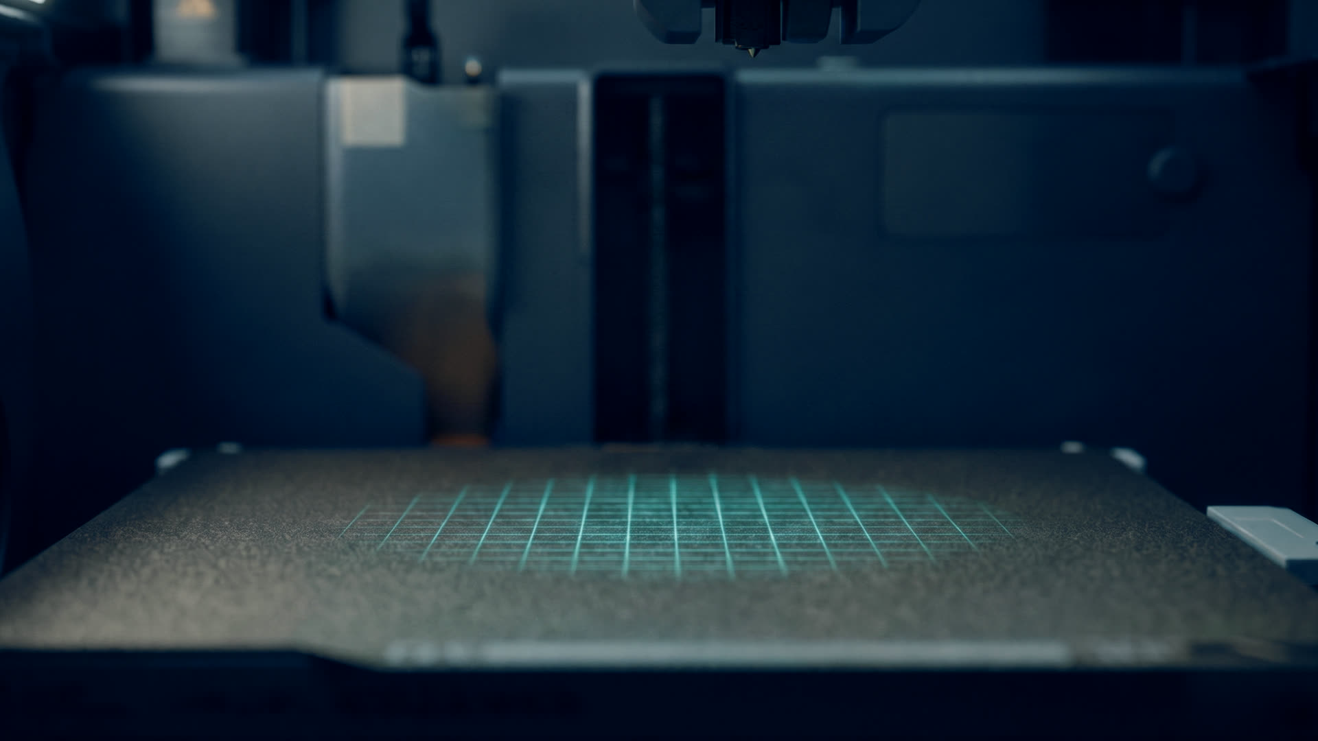
PrintGraft
Z angio-TK do szablonu aorty w minuty, nie w godziny. Autorskie oprogramowanie automatycznie segmentuje aortę pacjenta i przygotowuje model odcinka aorty do druku szablonu 3D. Hybrydowa architektura sieci neuronowych, 3D U-Net i trzy płaszczyznowe 2D U-Nety, trenowana na 222 badaniach angio-TK, uniezależnia wynik od operatora. Rdzeń technologiczny randomizowanego badania ABM. From CT angiography to an aortic template in minutes, not hours. Proprietary software automatically segments the patient's aorta and prepares the model of the aortic segment for 3D template printing. A hybrid neural-network architecture, a 3D U-Net and three plane-specific 2D U-Nets, trained on 222 CT angiographies, makes the result operator-independent. The technological core of the ABM randomized trial.
Segmentacja, która nie zależy od tego, kto siedzi przy komputerzeSegmentation that does not depend on who sits at the computer
Szablon 3D jest tak dobry, jak model, z którego powstał. Ręczna segmentacja aorty z angio-TK zajmuje godziny i różni się w zależności od osoby, która ją wykonuje. PrintGraft robi to automatycznie: sieć 3D U-Net odczytuje objętość badania, a trzy płaszczyznowe sieci 2D U-Net dopracowują granice naczynia w przekrojach osiowym, czołowym i strzałkowym. Zbiór treningowy to 222 badania angio-TK. A 3D template is only as good as the model it came from. Manual aortic segmentation from CT angiography takes hours and varies with the person doing it. PrintGraft does it automatically: a 3D U-Net reads the scan volume and three plane-specific 2D U-Nets refine the vessel boundaries in the axial, coronal and sagittal planes. The training set is 222 CT angiographies.
Wynikiem jest gotowy do druku model odcinka aorty z odejściami naczyń trzewnych. Ten model jest wspólnym mianownikiem dwóch technologii: w Metodzie Szczecińskiej zostaje wydrukowany jako fizyczny szablon, na którym chirurg modyfikuje stent-graft (PMEG), a w HoloGraft staje się hologramem w goglach mieszanej rzeczywistości. W wieloośrodkowym badaniu randomizowanym Agencji Badań Medycznych PrintGraft generuje szablony dla wszystkich ośrodków. The result is a print-ready model of the aortic segment with the visceral branch origins. This model is the common denominator of two technologies: in the Szczecin Method it is printed as a physical template on which the surgeon modifies the stent-graft (PMEG), and in HoloGraft it becomes a hologram in a mixed-reality headset. In the multicenter randomized trial of the Medical Research Agency, PrintGraft generates the templates for all centers.
Workflow w czterech krokachA four-step workflow
Badanie pacjenta trafia do PrintGraft w oryginalnej rozdzielczości; nie wymaga wstępnej obróbki.The patient's scan enters PrintGraft at its original resolution; no preprocessing is required.
3D U-Net wyznacza światło aorty w całej objętości, trzy 2D U-Nety dopracowują granice w każdej płaszczyźnie. Wynik nie zależy od operatora.The 3D U-Net delineates the aortic lumen across the volume, three 2D U-Nets refine the boundaries in each plane. The result is operator-independent.
Model jest przycinany do odcinka docelowego z odejściami naczyń i przygotowywany do druku 3D jako szablon aorty.The model is cropped to the target segment with the vessel origins and prepared for 3D printing as an aortic template.
Wydrukowany szablon trafia na salę: chirurg nakłada na niego stent-graft i wyznacza fenestracje. Pełny opis: Metoda Szczecińska.The printed template goes to the OR: the surgeon fits the stent-graft over it and marks the fenestrations. Full description: the Szczecin Method.
PrintGraft krok po krokuPrintGraft step by step
Od angio-TK do angiografii kontrolnej — jeden powtarzalny tor postępowania w dziewięciu scenach: segmentacja, szablon, druk, sterylizacja, modyfikacja stent-graftu na stoliku i implantacja. Rozdziały poniżej przewijają film do wybranego etapu.From CT angiography to completion angiography — one reproducible pathway in nine scenes: segmentation, template, printing, sterilization, back-table graft modification and implantation. The chapters below seek the video to any stage.
Transkrypcja narracjiNarration transcript
PrintGraft. Szablon do modyfikacji stent-graftu przez chirurga. Punktem wyjścia jest rutynowe angio-TK. Segmentacja aorty jest w pełni automatyczna, oparta na sieci neuronowej, bez ręcznej edycji konturów. Kontury składają się w model aorty. Usuwamy wszystko poza obszarem zainteresowania: aortę piersiową, aortę brzuszną z tętnicami biodrowymi oraz gałęzie trzewne i nerkowe, zostawiając przy każdym ujściu otwór prowadzący. Segment wydrążamy do ściany grubości 1,5 mm. Tak powstaje szablon. Szablon zapisujemy w formacie STL i przekazujemy bezpośrednio na drukarkę trójwymiarową. Druk SLA w żywicy biozgodnej, odpornej na tlenek etylenu. Sztywna skorupa segmentu trzewnego z otworem prowadzącym na każde naczynie docelowe. Sterylizacja tlenkiem etylenu w temperaturze 55 °C — niska temperatura nie odkształca szablonu. Stent-graft wysunięty z koszulki rozkładamy na jałowym stoliku. Szablon ustawiamy równo z proksymalnym brzegiem i przesuwamy, aż żadne przęsło stentu nie przecina otworu prowadzącego. Przez otwory prowadzące znaczymy miejsca fenestracji i zdejmujemy szablon. Każdą fenestrację wypalamy termokauterem pomiędzy przęsłami stentu, a następnie wzmacniamy pierścieniem nitinolowym. Przez fenestrację krezkową wprowadzamy prowadnik, po czym protezę ładujemy z powrotem do koszulki systemu dostarczającego w krymperze typu TAVI, nie opuszczając pola jałowego. Komponent fenestrowany rozprężamy w segmencie trzewnym z kontrolą rotacji. Naczynia docelowe kaniulujemy i mostkujemy stent-graftami rozflarowanymi wewnątrz protezy. Następnie wszczepiamy komponent aortalno-dwubiodrowy z odnogą długą i krótką, każdą przedłużoną stent-graftem biodrowym. Konstrukcja jest kompletna: część fenestrowana w segmencie trzewnym, poniżej rekonstrukcja aortalno-dwubiodrowa. Angiografia kontrolna — tętnica krezkowa górna i obie tętnice nerkowe drożne, worek tętniaka wyłączony z krążenia. Od tomografii do implantacji: jeden powtarzalny tor postępowania. PrintGraft.
PrintGraft, a template for physician modification of a stent-graft. The starting point is a routine CT angiogram. Segmentation of the aorta is fully automatic, performed by a neural network with no manual editing of the contours. The contours form a 3D model of the aorta. Everything outside the region of interest is removed: thoracic aorta, abdominal aorta with the iliac arteries, and the side branches, leaving a guide hole at each ostium. The remaining segment is hollowed into a 1.5 mm wall. This is the template. The template is saved as an STL file and sent directly to the 3D printer. Printed by stereolithography in a biocompatible, ethylene-oxide-tolerant resin. A rigid shell of the visceral aorta with one guide hole per target vessel. Sterilization with ethylene oxide at 55 °C — the low temperature leaves the template undistorted. The stent-graft withdrawn from its sheath is laid out on the sterile back table. The template is aligned with the proximal fabric edge and shifted until no stent strut crosses a guide hole. The fenestration sites are marked through the guide holes, and the template is removed. Each fenestration is burned open with a thermocautery between the stent struts and reinforced with a nitinol ring. A guide wire is passed through the SMA fenestration, and the graft is loaded back into the delivery system sheath in a TAVI-type crimper within the sterile field. The fenestrated component is deployed in the visceral segment under rotational control. The target vessels are cannulated and bridged with covered stents flared inside the graft. An aorto-bi-iliac component follows with a long and a short limb, each extended with an iliac stent-graft. The construct is complete: the fenestrated segment above, the aorto-bi-iliac reconstruction below. Completion angiography — the superior mesenteric artery and both renal arteries are patent, the aneurysm sac is excluded. From CT to implantation, one reproducible pathway. PrintGraft.
Dowody, nie deklaracjeEvidence, not claims
Szablon aorty zwalidowany krok po krokuThe aortic template validated step by step
Najczęstsze pytaniaFrequently asked questions
Co robi PrintGraft?What does PrintGraft do?
Automatycznie segmentuje aortę z badania angio-TK i przygotowuje model odcinka aorty do druku szablonu 3D. Zastępuje wielogodzinną segmentację ręczną: szablon jest gotowy w minuty, niezależnie od operatora.It automatically segments the aorta from CT angiography and prepares the model of the aortic segment for 3D template printing. It replaces hours of manual segmentation: the template is ready in minutes, operator-independently.
Jak zbudowane są sieci neuronowe PrintGraft?How are PrintGraft's neural networks built?
To architektura hybrydowa: sieć 3D U-Net oraz trzy płaszczyznowe sieci 2D U-Net, trenowane na 222 badaniach angio-TK.It is a hybrid architecture: a 3D U-Net and three plane-specific 2D U-Nets, trained on 222 CT angiographies.
Jak PrintGraft łączy się z Metodą Szczecińską i HoloGraft?How does PrintGraft relate to the Szczecin Method and HoloGraft?
PrintGraft dostarcza cyfrowy model aorty. W Metodzie Szczecińskiej model jest drukowany jako fizyczny szablon, na którym chirurg modyfikuje stent-graft (PMEG). W HoloGraft ten sam model staje się hologramem w goglach mieszanej rzeczywistości.PrintGraft provides the digital model of the aorta. In the Szczecin Method the model is printed as a physical template on which the surgeon modifies the stent-graft (PMEG). In HoloGraft the same model becomes a hologram in a mixed-reality headset.
Gdzie PrintGraft jest używany?Where is PrintGraft used?
Jest rdzeniem technologicznym wieloośrodkowego badania randomizowanego Agencji Badań Medycznych porównującego PMEG na szablonie 3D z protezami fabrycznymi (NCT06969729; ośrodki: USK-2 PUM jako koordynator, UM Poznań, 4. WSK Wrocław).It is the technological core of the Medical Research Agency's multicenter randomized trial comparing PMEG on a 3D template with factory-made prostheses (NCT06969729; centers: USK-2 PUM as coordinator, Poznań University of Medical Sciences, 4th Military Clinical Hospital in Wrocław).